
Clinical assessment of the knee joints in dogs (PL)
Patellar luxation is the pathological displacement of the patella (kneecap) from its correct anatomical position in the intercondylar (trochlear) groove of the femur.
Contents
In a healthy joint, the patella glides steadily and precisely along the intercondylar groove, ensuring optimal biomechanics of the knee joint. When this mechanism is disrupted, the patella becomes displaced, which may be accompanied by changes in gait, reduced function of the limb and the onset of pain.
Accurate diagnosis and professional assessment of the knee joints are essential for maintaining the animal’s health and for making well-founded decisions in breeding.
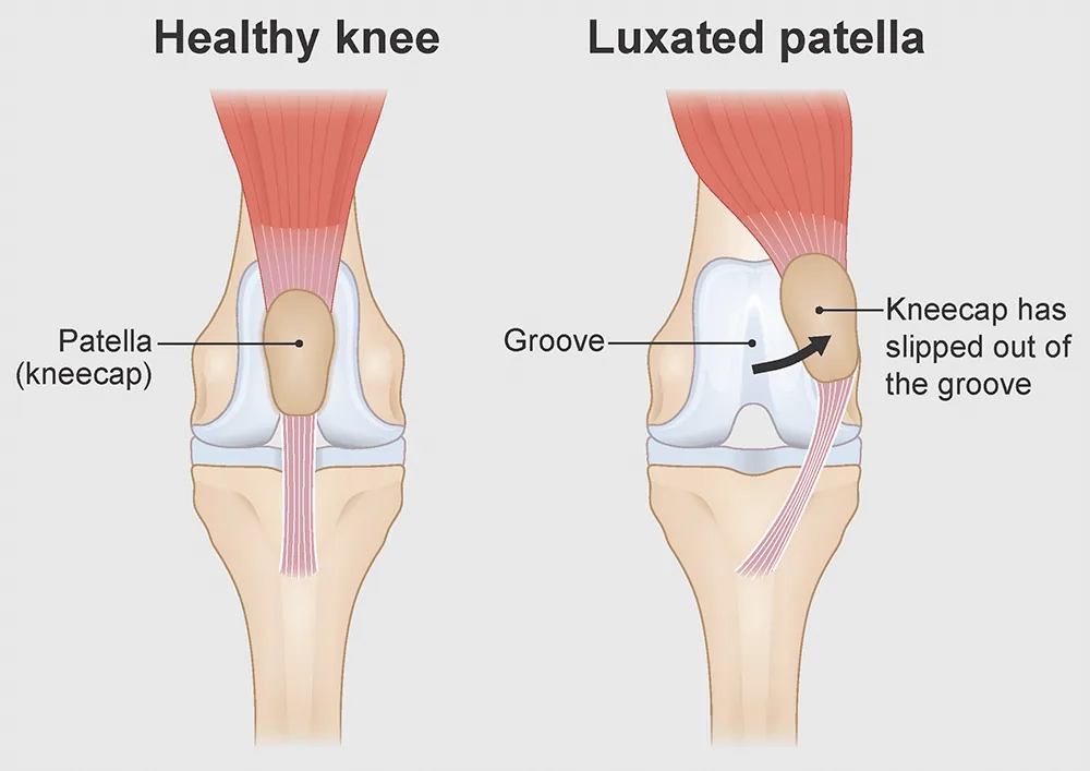
- Healthy knee – a healthy knee joint
- Luxated patella – patellar luxation
- Patella (kneecap) – the patella
- Groove – the intercondylar (trochlear) groove
- Kneecap has slipped out of the groove – displacement of the patella from its normal position in the femoral trochlea
Regulations on the clinical assessment of the knee joints (PL)
International certificate of clinical assessment of the knee joints (PL)
Patellar luxation grade (PL)
The patellar luxation grade (PL) is given in accordance with the classification adopted by the FCI.
For the knee joints
- PL-0: No patellar luxation. The knee joint is clinically healthy.
- PL-1: The patella can be displaced manually with the knee joint in extension (straightened), after which it spontaneously returns to its correct anatomical position.
- PL-2: The patella luxates when the knee joint is flexed (bent), when the lower leg is rotated or on manual manipulation, and does not spontaneously return to its correct anatomical position.
- PL-3: Permanent patellar luxation; the patella can be manually repositioned in its correct anatomical position, but the luxation recurs.
- PL-4: Fixed patellar luxation in which manual repositioning is impossible.
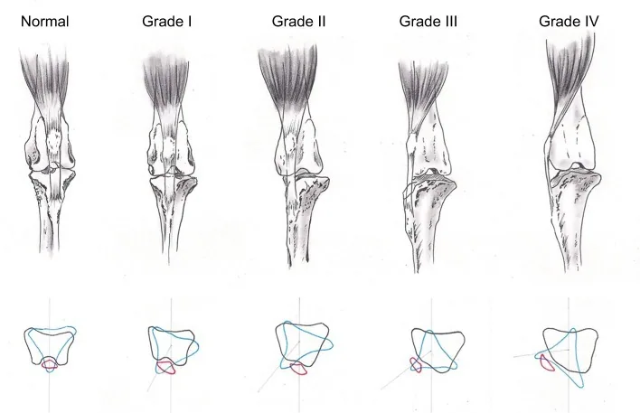
Schematic illustration of the patellar luxation grades (PL)
The image compares the normal position of the patella with the different grades of luxation (PL I–IV) in a cranial view.
In a healthy joint, the axis of the quadriceps femoris mechanism runs centrally along the femur, keeping the patella stably positioned in the trochlear (intercondylar) groove and ensuring optimal biomechanics of the knee joint.
- In grade PL I, the patella can be pushed out of the groove by manual manipulation and then spontaneously returns to its correct anatomical position.
- In grade PL II, the patella becomes displaced during movement or under external force and may stay out of place without spontaneously returning, reflecting an early loss of stability of the extensor mechanism.
- In grade PL III, the patella is permanently luxated; it can be temporarily repositioned by hand, but the luxation quickly recurs because of a marked deviation of the axis of the extensor mechanism.
- In grade PL IV, the patella lies permanently outside the trochlear groove and cannot be repositioned by hand; there are significant structural changes, including bone deformities and flattening of the groove.
The lower diagram shows the progressive shift of the axis of the extensor mechanism and the rotation of the tibia relative to the femur, which underlie the change in the position of the patella.
Veterinarians accredited for PL assessment
Veterinarians accredited by the Cynological Union of Moldova and authorised to carry out the clinical assessment of the knee joints in dogs (PL):
Our contacts
- Address
- 7 Alexandru cel Bun St., office 208, Chișinău
- Monday – Thursday
- 09:00–18:00
- Friday
- 09:00–17:00
- Saturday, Sunday
- Closed
- Phone
- +373 79 170 889
- info@chinolog.md