
X-ray examination of the hip (HD) and elbow (ED) joints in dogs
Dysplasia is a disorder of the growth and development of the joints that impairs their mobility and causes structural changes in the form of damage to parts of the joint and degenerative joint changes.
Contents
- The hip joint
- The elbow joint
- Regulations on hip (HD) and elbow (ED) dysplasia screening
- International certificate for hip (HD) and elbow (ED) dysplasia
- Information leaflets (British Veterinary Association)
- Hip (HD) and elbow (ED) dysplasia grades
- Specialists accredited by the Cynological Union of Moldova
- FCI information on hip and elbow dysplasia
- Our contacts
Analysing X-rays of a dog’s hip and/or elbow joints makes it possible to detect signs of disease in these joints (dysplasia) and to prevent animals found to have such conditions from being used for breeding, thereby stopping the genetic spread of the disease.
The hip joint
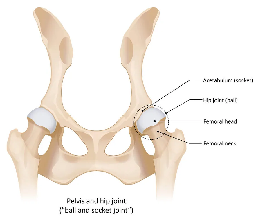
- Acetabulum (socket) – the acetabulum (cotyloid cavity)
- Hip joint (ball) – the hip (coxofemoral) joint
- Femoral head – the head of the femur
- Femoral neck – the neck of the femur
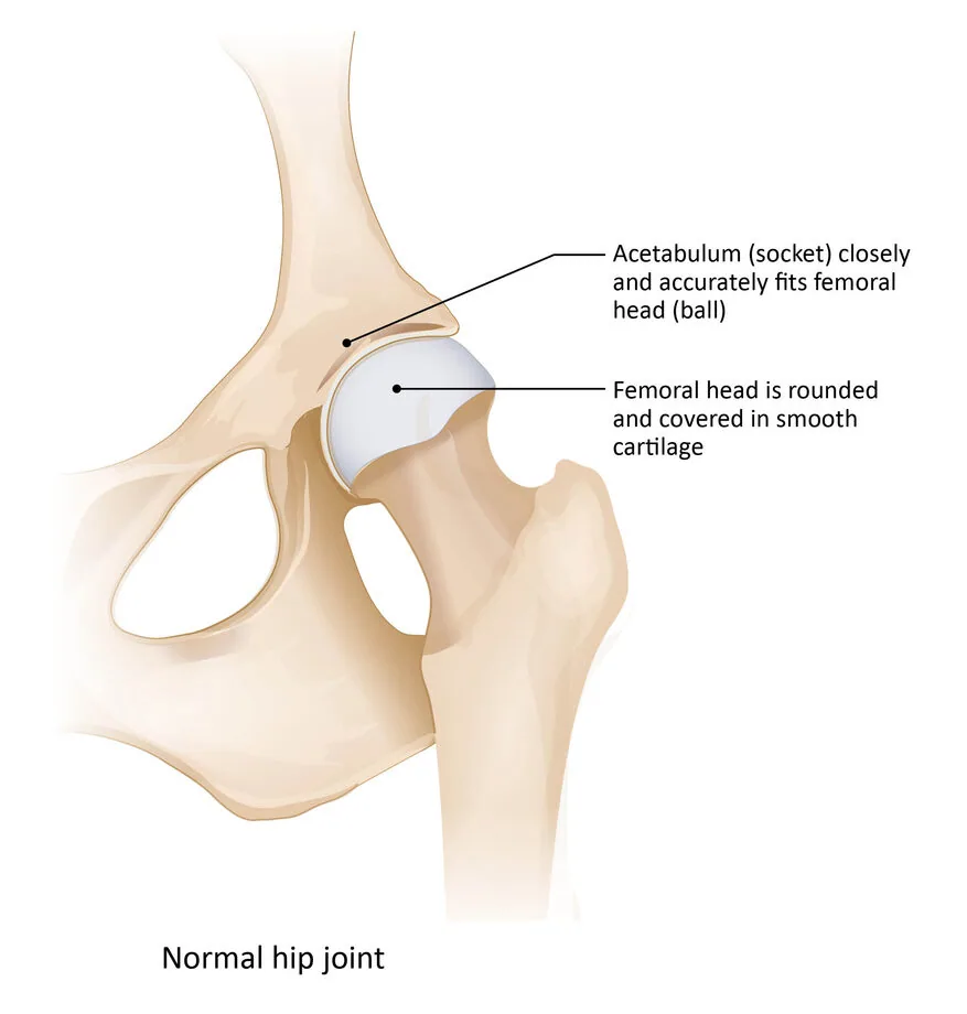
- Acetabulum (socket) closely and accurately fits femoral head (ball) – the acetabulum fits the femoral head snugly and precisely
- Femoral head is rounded and covered in smooth cartilage – the femoral head is round and covered with smooth articular cartilage
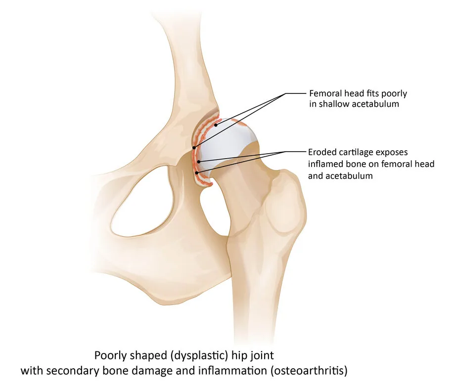
- Femoral head fits poorly in shallow acetabulum – the femoral head does not sit snugly enough in the shallow acetabulum
- Eroded cartilage exposes inflamed bone on femoral head and acetabulum – damage to the articular cartilage exposes inflamed areas of bone on the femoral head and the acetabulum
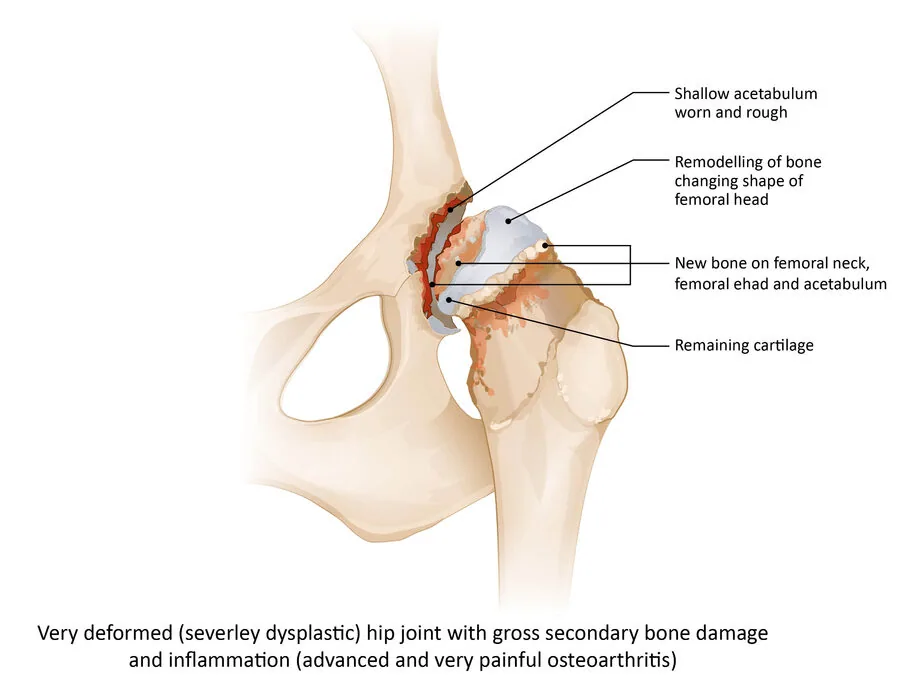
- Shallow acetabulum worn and rough – a shallow acetabulum with a worn, uneven articular surface
- Remodelling of bone changing shape of femoral head – bone remodelling that alters the shape of the femoral head
- New bone on femoral neck, femoral head and acetabulum – newly formed bone on the femoral neck and head and on the acetabulum
- Remaining cartilage – the articular cartilage that is still intact
The elbow joint
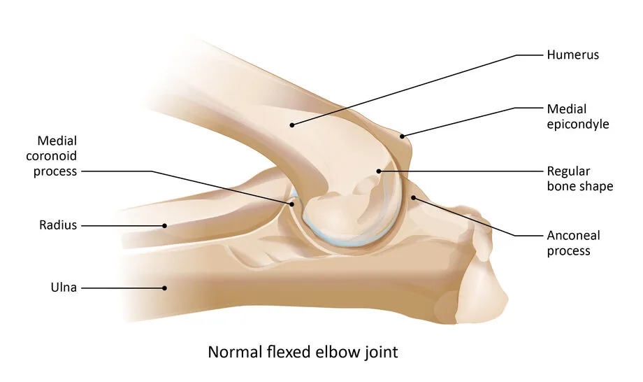
- Humerus – the upper arm bone
- Medial epicondyle – the medial epicondyle of the humerus
- Regular bone shape – normal bone shape
- Anconeal process – the anconeal process of the ulna
- Medial coronoid process – the medial coronoid process of the ulna
- Radius – the radius
- Ulna – the ulna
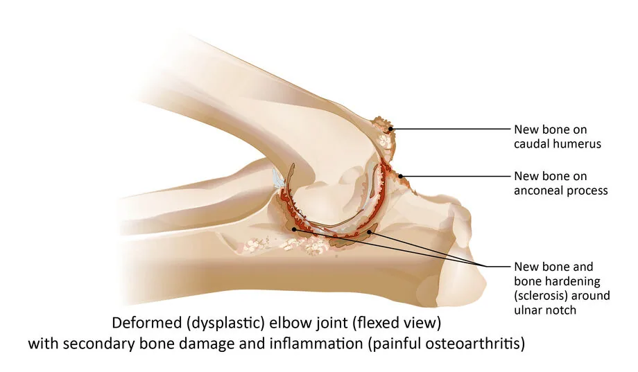
- New bone on caudal humerus – newly formed bone on the caudal surface of the humerus
- New bone on anconeal process – newly formed bone on the anconeal process
- New bone and bone hardening (sclerosis) around ulnar notch – newly formed bone and bone sclerosis around the trochlear notch of the ulna
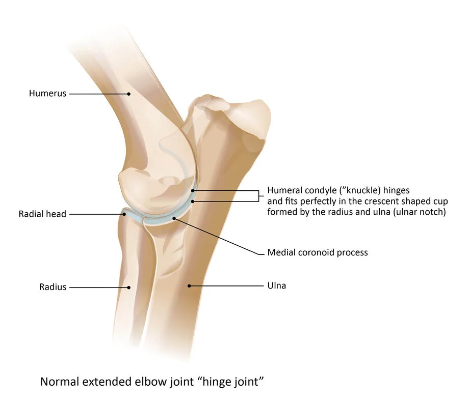
- Humerus – the upper arm bone
- Radial head – the head of the radius
- Radius – the radius
- Ulna – the ulna
- Medial coronoid process – the medial coronoid process of the ulna
- Humeral condyle (“knuckle”) hinges and fits perfectly in the crescent shaped cup formed by the radius and ulna (ulnar notch) – the humeral condyle forms a hinge and matches exactly the crescent-shaped articular surface formed by the radius and ulna (the trochlear notch of the ulna)
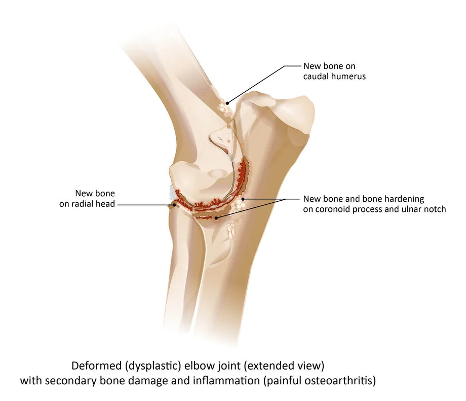
- New bone on caudal humerus – newly formed bone on the caudal surface of the humerus
- New bone on radial head – newly formed bone on the head of the radius
- New bone and bone hardening on coronoid process and ulnar notch – newly formed bone and bone sclerosis in the area of the coronoid process and the trochlear notch of the ulna
Regulations on hip (HD) and elbow (ED) dysplasia screening
International certificate for hip (HD) and elbow (ED) dysplasia
Information leaflets (British Veterinary Association)
Hip (HD) and elbow (ED) dysplasia grades
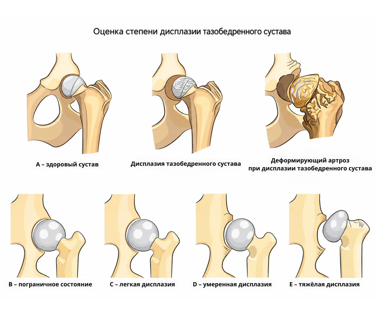
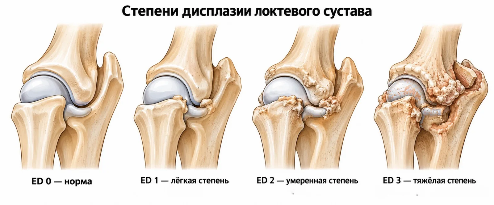
The severity of dysplasia is graded according to the classification adopted by the FCI.
The overall grade is assigned on the basis of the least favourable result.
For the hip joints
- A – healthy joint;
- B – borderline;
- C – mild dysplasia;
- D – moderate dysplasia;
- E – severe dysplasia.
For the elbow joints
- 0 – healthy joint;
- 1 – mild osteoarthritis;
- 2 – moderate osteoarthritis;
- 3 – severe osteoarthritis or a primary lesion.
Specialists accredited by the Cynological Union of Moldova
Veterinary clinic accredited by the Cynological Union of Moldova and authorised to carry out X-ray examination of the hip (HD) and elbow (ED) joints in dogs:
Veterinarian accredited by the Cynological Union of Moldova and authorised to carry out X-ray examination of the hip (HD) and elbow (ED) joints in dogs:
- Antonina Dumitriu (PhD in veterinary sciences)
Veterinarians accredited by the Cynological Union of Moldova and authorised to evaluate X-rays of the hip (HD) and elbow (ED) joints in dogs:
FCI information on hip and elbow dysplasia
- PDFFCI Requirements for Official Hip Dysplasia Screening
- PDFRadiographic Procedure for Hip Dysplasia Evaluation
- PDFFCI HIP Dysplasia Classification
- PDFHip-/Elbow Dysplasia International Certificate
- URLElbow dysplasia (ED) (Margarita Durán)Presentation
Our contacts
- Address
- 7 Alexandru cel Bun St., office 208, Chișinău
- Monday – Thursday
- 09:00–18:00
- Friday
- 09:00–17:00
- Saturday, Sunday
- Closed
- Phone
- +373 79 170 889
- info@chinolog.md